Retinal Tear Optos - Retinal Detachment Recognizing Pathology Optos / An optos® retinal exam provides:. Retinal defects come in different shapes and sizes and may be either partial or full thickness. Retinal holes and tears are commonly encountered during dilated fundus examination of both symptomatic and asymptomatic patients. It is important to detect problems in the eye, like diabetes, macular degeneration, melanoma, bleeding, hypertensive retinopathy and glaucoma before they cause loss of vision. But, diseases such as macular degeneration, glaucoma, retinal tears or detachments, and other health problems such as diabetes and high blood pressure can be seen with a thorough exam of the retina. The most common reason for a rd is a retinal tear and the most common reason for a tear is a pvd.
Our astoria optometrists are able to monitor freckles in the back of the eye known as a choroidal nevus. The optomap® retinal exam produces an image that is as unique as you fingerprint and provides us with a wide view to look at the health of your retina. The retina is the part of your eye that captures the image of what you are looking at, similar to film in a camera. A retinal tear, or retinal detachment, occurs when the retina is torn from the underlying tissue. The device allows for the discovery of retinal melanomas and.
Flashes Floaters from skopticians.co.uk This is likely due to the vessels blocking the exiting light. A retinal tear, or retinal detachment, occurs when the retina is torn from the underlying tissue. Retinal imaging takes a digital picture of the back of your eye. Optos retinal imaging the optos imaging technology allows us to give our patients a more thorough health evaluation. Our astoria optometrists are able to monitor freckles in the back of the eye known as a choroidal nevus. A retinal detachment (rd) is the separation of the sensory retina from the retinal pigment epithelium (rpe) (outer segments of the photoreceptors from the microvilli of the rpe). A scan to show a healthy eye or detect disease. An optomap® retinal exam provides:
But, diseases such as macular degeneration, glaucoma, retinal tears or detachments, and other health problems such as diabetes and high blood pressure can be seen with a thorough exam of the retina.
Jason calhoun, mayo clinic jacksonville, department of ophthalmology. Optos retinal imaging the optos imaging technology allows us to give our patients a more thorough health evaluation. This combined device facilitates the early detection, management and effective treatment of disorders and diseases evidenced in the retina such as retinal detachments and tears, glaucoma, diabetic retinopathy. A scan to show a healthy eye or detect disease. Our astoria optometrists are able to monitor freckles in the back of the eye known as a choroidal nevus. An optomap® retinal exam provides: A giant retinal tear typically develops slightly posterior and parallel to the ora serrata, with the vitreous base attached to the anterior margin of the tear. But, diseases such as macular degeneration, glaucoma, retinal tears or detachments, and other health problems such as diabetes and high blood pressure can be seen with a thorough exam of the retina. Retinal tears when a retinal tear or hole hasn't yet progressed to detachment, your eye surgeon may suggest one of the following procedures to prevent retinal detachment and preserve vision. His idea came to fruition, and optos retinal imaging quickly became a coveted method of examining the retina. Retinal tear temporal about 4 o'clock in the left eye. We offer the optomap® retinal exam as an important part of our temple eye exams. But, diseases such as macular degeneration, glaucoma, retinal tears or detachments, and other health problems such as diabetes and high blood pressure can be seen with a thorough exam of the retina.
Retinal tears when a retinal tear or hole hasn't yet progressed to detachment, your eye surgeon may suggest one of the following procedures to prevent retinal detachment and preserve vision. A scan to show a healthy eye or detect disease. Retinal holes and tears are commonly encountered during dilated fundus examination of both symptomatic and asymptomatic patients. Some times the retinal vessels on the detachment will be dark in appearance, as seen in a retinoschisis. Optos retinal exam annual eye exams are vital to maintaining your vision and overall health.
Retinal Detachment Right Eye Optomap Retina Image Bank from imagebank.asrs.org A retinal tear, or retinal detachment, occurs when the retina is torn from the underlying tissue. An optomap® retinal exam provides: The device allows for the discovery of retinal melanomas and. His idea came to fruition, and optos retinal imaging quickly became a coveted method of examining the retina. Optos retinal exam annual eye exams are vital to maintaining your vision and overall health. The most common reason for a rd is a retinal tear and the most common reason for a tear is a pvd. Tearing of the retina may produce retinal or vitreous hemorrhages. But, diseases such as macular degeneration, glaucoma, retinal tears or detachments, and other health problems such as diabetes and high blood pressure can be seen with a thorough exam of the retina.
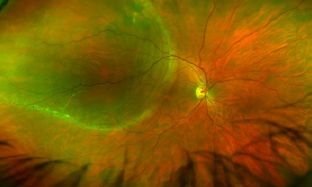
Retinal tear temporal about 4 o'clock in the left eye.
Their causes are equally varied. Another condition leading to retinal detachment is a giant retinal tear, defined as a retinal tear that extends 3 clock hours (90 degrees) or more around the circumference of the globe. This combined device facilitates the early detection, management and effective treatment of disorders and diseases evidenced in the retina such as retinal detachments and tears, glaucoma, diabetic retinopathy. A retinal tear, or retinal detachment, occurs when the retina is torn from the underlying tissue. It shows the retina (where light and images hit), the optic disk (a spot on the retina that holds the optic nerve, which sends. The most frequent cause of an operculated tear is a pvd. An optomap® retinal exam provides: Retinal tear temporal about 4 o'clock in the left eye. The most frequent cause of an operculated tear is a pvd. A scan to show a healthy eye or detect disease. A retinal detachment (rd) is the separation of the sensory retina from the retinal pigment epithelium (rpe) (outer segments of the photoreceptors from the microvilli of the rpe). This advancement allows doctors to see 82 percent of the retina in a single capture, and. Jason calhoun, mayo clinic jacksonville, department of ophthalmology.
She immediately followed up with a retinal specialist who performed an emergency laser surgery that successfully repaired the tear. This is likely due to the vessels blocking the exiting light. A retinal detachment (rd) is the separation of the sensory retina from the retinal pigment epithelium (rpe) (outer segments of the photoreceptors from the microvilli of the rpe). But, diseases such as macular degeneration, glaucoma, retinal tears or detachments, and other health problems such as diabetes and high blood pressure can be seen with a thorough exam of the retina. The optomap® retinal exam produces an image that is as unique as you fingerprint and provides us with a wide view to look at the health of your retina.
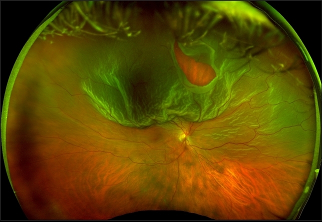
Optos Giant Tear Within Retinal Detachment Retina Image Bank from imagebank.asrs.org A retinal tear, or retinal detachment, occurs when the retina is torn from the underlying tissue. But, diseases such as macular degeneration, glaucoma, retinal tears or detachments, and other health problems such as diabetes and high blood pressure can be seen with a thorough exam of the retina. The most frequent cause of an operculated tear is a pvd. An optomap® retinal exam provides: +61 8 8444 6500 auinfo@optos.com building the retina company optos.com opto map color image highlighting both the retinal tear and pvd, both in the far periphery. Optos retinal photography many eye problems can develop without you knowing, in fact, you may not even notice any change in your sight, diseases or damage such as macular degeneration, glaucoma, retinal tears or detachments, and other health problems such as diabetes and high blood pressure can be seen with a thorough exam of the retina. Retinal defects come in different shapes and sizes and may be either partial or full thickness. An optos® retinal exam provides:
The most frequent cause of an operculated tear is a pvd.
+61 8 8444 6500 auinfo@optos.com building the retina company optos.com opto map color image highlighting both the retinal tear and pvd, both in the far periphery. The most frequent cause of an operculated tear is a pvd. The optomap® retinal exam produces an image that is as unique as you fingerprint and provides us with a wide view to look at the health of your retina. This advancement allows doctors to see 82 percent of the retina in a single capture, and. Retinal imaging takes a digital picture of the back of your eye. Another condition leading to retinal detachment is a giant retinal tear, defined as a retinal tear that extends 3 clock hours (90 degrees) or more around the circumference of the globe. The retina is the part of your eye that captures the image of what you are looking at, similar to film in a camera. She immediately followed up with a retinal specialist who performed an emergency laser surgery that successfully repaired the tear. An optomap® retinal exam provides: Their causes are equally varied. If you think that you are suffering from a retinal tear, please call your optometrists at the vision center in fort worth and lubbock, tx. A giant retinal tear typically develops slightly posterior and parallel to the ora serrata, with the vitreous base attached to the anterior margin of the tear. Our astoria optometrists are able to monitor freckles in the back of the eye known as a choroidal nevus.
We offer the optomap® retinal exam as an important part of our temple eye exams retinal tear. This is likely due to the vessels blocking the exiting light.


0 Komentar